Ophthalmology Services

Treating Eye Problems in Pets
Eye health and effective vision are very important to your pets' quality of life. During regular checkups, your primary care veterinarian will monitor your pets' eye health, as part of a general assessment.
If your pet has a minor ocular condition or injury requiring treatment, in many cases your veterinarian will be handling it. For more complex cases, however, you may be referred to or seek assistance of a board-certified veterinary ophthalmologist who has specialized training, experience and equipment. Veterinary ophthalmologists are veterinarians who undergo three to four additional years of training in an approved residency program, and specialize exclusively on medical and surgical treatment of the animal eye. At the end of the residency, they must complete the board certification process, involving a three-day exam consisting of written, practical and surgical portions.
All board-certified veterinary ophthalmologists are Diplomates of the American College of Veterinary Ophthalmologists (DACVOs). The ACVO is a specialty organization affiliated with the American Veterinary Medical Association.
To learn more about the American College of Veterinary Ophthalmologists, please visit the ACVO website. www.acvo.org
The Ophthalmology Service at Tufts VETS offers the most advanced diagnostic and treatment practices for eye diseases and injuries in small animals. We provide clear and complete explanations of the problem and the appropriate treatment for both you and your veterinarian. We strive to deliver optimum medical care which will provide the best possible outcome for your pet.

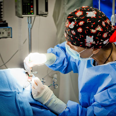
Surgical Services
Eyelid Surgery
- Entropion/Ectropion
- Correction of macropalpebral fissure (brachycephalic breeds, such as Shih Tzu, Pekingese, Pug)
- Laceration repair
- Simple eyelid tumor removal with liquid nitrogen cryotreatment
- Reconstructive palpebral surgery for extensive defect/tumors
- Distichiasis (abnormal eyelashes) cryotreatment/thermal electrocautery
- Ectopic cilia permanent removal
- Hyaluronan eyelid filler injection for entropion treatment
Third Eyelid Surgery
- Third eyelid gland prolapse surgical treatment ("cherry eye" repositioning)
- Third eyelid cartilage eversion/inversion permanent correction
Corneal Surgery
- Laceration and perforation repair
- Full penetrating or lamellar corneal grafting
- Conjunctival graft
- Grid keratotomy
- Diamond Burr Superficial Keratectomy (DBSK)
- Superficial keratectomy
- Corneal and corneoscleral tumors removal
- Dermoid removal
- Corneal sequestrum removal
- Cryotreatment of pigmentary keratitis
Cataract Evaluation & Surgery
We perform cataract surgery through phacoemulsification and IOL (artificial Intra-Ocular Lens) implantation. We insert foldable IOLs that provide your pet with the least invasive and inflammatory procedure available today. This type of surgery is the same as the surgery performed in people, the outcome of which is reported to be the most successful. Anterior vitrectomy is also available if the pet requires it. A video about cataract surgery can be viewed here.
Lens Instability Surgery
Lens instability is a severe concern in specific breeds, such as Terriers and Terrier crosses, which are predisposed to its development. Anterior lens luxation is considered a surgical emergency and requires immediate attention. According to the specific case, intracapsular lens removal or manual repositioning may be recommended.
Uveal Procedures
- Transcorneal photocoagulation (laser) for treatment of uveal melanomas or uveal cysts
- Retinopexy for partial retinal detachment
Glaucoma Procedures
- Endoscopic cyclophotocoagulation (“endolaser”) for Glaucoma Treatment
This sophisticated piece of equipment allows the newest, most advanced surgical treatment for glaucoma. We are currently among the few practices in New England to offer this procedure, which controls and treats glaucoma. Presurgical evaluation and clinical assessment will be required. A small goniodevice (silicone valve) may be also implanted according to the specific case.
End Stage Procedures
Sometimes despite all our efforts or due to severe damage to the eye or the presence of intraocular tumors, the best surgical option for your pet is the removal of the eye. This can be performed through a simple procedure (enucleation), with or without orbital prosthetics, or, if applicable, through a more complex procedure with an intraocular prosthetic implantation. During enucleation, a long-acting anesthetic is injected in the orbit, guaranteeing your pet’s immediate wellbeing and comfort.
*** Please note that a complete physical exam and blood testing are required before surgery.
Common Ocular Conditions
- Eyelash/eyelid abnormalities
- Dry eye
- Allergic/immune-mediated conjunctivitis
- Superficial and deep corneal ulcers
- Cataracts
- Glaucoma
- Uveitis
- Retinal detachment/ retinal degeneration
- Eyelid and intraocular tumors
Symptoms to Watch for and Report to Your Veterinarian
- Squinting and tearing
- Ocular discharge
- Red eye or ocular discoloration
- Cloudy eye
- Decreased vision
- Rapidly growing or unusual eyelid masses
Complete Ophthalmology Services
Diagnosing and treating eye disorders can involve both simple and more advanced methods. Tufts VETS offers you and your veterinarian a full range of diagnostic and surgical services.
Routine Ophthalmic Examination
The initial diagnostics that are done on almost all veterinary patients will include a Schirmer Tear Test / Strip Meniscometry Test, corneal staining, intraocular pressure assessment, pupillary dilation.
A Schirmer Tear Test measures the eye’s tear production. Veterinarians will perform this test anytime the eye is red and has discharge. The test is performed by taking a tear test strip and placing the end between the lower eyelid and eye. The amount of moisture wicking up the strip in a minute is measured. Normal values vary between species but for the dog 20 mm +/- 5 mm in 1 minute is considered normal. A biomicroscope, a specialized piece of equipment provided with focal light source and magnification is then used to examine the eyelids, structures surrounding the eye, and front half of the eye.
Vision is assessed through visual testing, including a menace response, the Pupillary Light Reflex (PLR) and a dazzle reflex.
The cornea is then stained with fluorescein dye. The dye will adhere to corneal structures if there is a break in the cornea’s epithelial surface and indicating an ulcer is present.
Intraocular pressure (IOP) is measured with a rebound tonometer. An elevated IOP may indicate glaucoma. One of the first signs of glaucoma may be redness and discharge so measuring IOP is an important diagnostic test to prevent elevated IOP from blinding an eye. Once intraocular pressures are determined to be within normal limits the pupils are dilated. Typically, dilation will take 10 to 20 minutes to complete, with pupils remaining dilated for a few hours.
Dilation of the pupil also allows a thorough examination of the lenses, through the use of the slit lamp or biomicroscope. The lens is assessed for its position, presence of opacities (cataracts) and presence of congenital defects.
Finally, indirect ophthalmoscopy is performed to examine the back or fundus of the eye (retina, choroid and optic nerve). When examining the fundus, the specialist will be looking at the number and character of blood vessels, size and shape of the optic nerve, pigmentation or depigmentation, retinal anatomy, and intensity of tapetal reflectivity (the tapetum is the part of the back of the animal’s eye that reflects light back out of the eye).
Additional Diagnostic Services
- Gonioscopy (to test risk of glaucoma)
- Cytology & Biopsy (conjunctival & corneal scrapings, fine needle aspirates, incisional or excisional biopsies - to identify infectious, inflammatory or neoplastic disorders)
- Macro pictures and fundus photography
- Ocular Ultrasonography (to evaluate ocular injuries, intraocular tumors, cataracts, retinal detachment & orbital diseases)
- CT scan and contrast radiography for intracranial and orbital disorders
- Doppler Systemic Blood Pressure Measurement (early recognition of systemic hypertension helps prevent & treat chorio-retinopathy and retinal detachment)
- Electroretinography (to assess retinal function)
- CAER/CERF Evaluations (to certify & register a dog free of heritable eye disease with the Canine Eye Animal Registry)

